The content is published under Creative Commons Attribution 4.0 International license.
Reviewed Article:
The Mummification of Votive Birds: Past and Present

A mummy is defined as a ‘well-preserved dead body’ (Cockburn, Cockburn & Reyman 1998, 1), achieved by either natural or anthropogenic methods and refers to both human and animal subjects. Mummies achieved through both these methods are found in ancient Egypt as a result of preservation through desiccation, achieved by direct contact between the corpse and a dry, sandy matrix (natural); or through the use of natron (anthropogenic), coupled with evisceration (the removal of the internal organs) and anointment with resinous compounds, followed by wrapping the corpse in layers of linen (Ikram and Dodson 1998; Taylor 2001).
Introduction
Animal mummies can be divided into four types: pets, mummified to accompany their owners into the Afterlife; victual, prepared and mummified food offerings for consumption in the Afterlife; cult, an individual selected based upon specific characteristics and markings to be an avatar for their prospective deity; and votive, where the whole population of certain species associated with a particular deity were classed as sacred and therefore worthy of mummification (Ikram and Iskander 2002; Ikram 2005; McKnight 2010).
Votive offerings comprised a wide variety of species including both domestic and wild mammals, fish, birds, reptiles and insects. Their purpose is uncertain, although it is thought that they acted as ‘go-between’ for deities and pilgrims during the Late-Roman Periods (664BC-AD395) at Saqqara (Ikram 2005, 9); in addition to their mummification and burial re-enacting the religious cycle of renewal and rebirth, which in turn ensured the political and economic stability of Egypt during its occupation by foreign rulers (Kessler and Nur el-Din 2005, 127-9; Von den Driesch et al 2006, 241). Evidence for specific materials and methods used in animal mummification are primarily focused on the mummified remains. Literary evidence for human mummification processes exist in ancient papyri and classical literature, alongside the Apis Embalming Papyrus, the surviving parts of which focus primarily on the wrapping techniques of the bulls sacred to the god Apis (Vos 1992). However, to date there is no surviving literary evidence referring to the mummification of votive animals in ancient Egypt. As a result of this, the mummies themselves remain the primary source of information (See Figure 1). This research focuses on bird remains as they comprise one of the largest proportions of animal mummies found from ancient Egypt.
Methods of bird mummification
For the most part birds were votive offerings and their mummification method has been described as a relatively simple procedure comprising desiccation and anointment techniques. Evisceration was thought to have been replaced by an internal douche, based on archaeological findings of enemas; followed by the use of natron as the desiccant, prior to being anointed with a resinous substance and wrapped in linen (Ikram 2005, 20-1). Research carried out by the Ancient Egyptian Animal Bio Bank team aimed to test such observations in the bird mummies studied in various museum collections using imaging (clinical X-ray and CT) and light microscopy with a particular focus on the following:
- Evidence for the use of evisceration;
- Evidence for the method of anointment;
- Evidence for the cause of death;
- Evidence for the use of an artificial desiccant, such as natron.
Radiographic observations
A lack of evisceration was observed (McKnight and Atherton in press) with several complete and articulated bird mummies identified as small birds of prey, mostly Falco tinnunculus (Kestrel) and Accipiter nisus (Sparrowhawk). The internal organs were thought to have become dehydrated and, as a result, had decreased in size within the abdominal cavity, and appeared as radiopaque shadows on radiographic images (See Figure 2).
Anointment methods, attributed to resinous substances that adhered to the bird during mummification, were observed as radiodense areas. These were visible in different ways: as a complete coating around the corpse (see Figure 3); or in patches, indicative of the resin1 settling in a particular area, synonymous with the final resting position, within the wrappings before becoming completely dry (Atherton 2012; McKnight 2010) (See Figure 4).
The presence of fractures as the cause of death was, in some cases, possible, particularly with significant cranial trauma; in addition to fractures to the cervical spine being previously attributed to over-rotation (Armitage and Clutton-Brock 1981). However, the frequent and rough post-mortem handling expected to have occurred during mummification and post-excavation, means that such diagnoses cannot be determined with any certainty. The authors hypothesise that perhaps such cervical fractures were the result of ‘rough-handling’ and significant amounts of tight linen wrappings in this region.
Microscopic observations
Light microscopy was useful in determining the absence of natron on feather samples (See Figure 5) (Atherton 2012); although linen samples from a bird mummy, thought to contain an ibis, from the Roman Period cemetery at Abydos showed that a natron ‘fingerprint’ was visible through the use of Environmental Scanning Electron Microscopy coupled with Electron Dispersive Spectroscopy (ESEM+EDS). It was hypothesised that this example demonstrated evidence for natron-soaked linen applied as the first layer of wrappings rather than the direct treatment of the bird corpse. The fact that some bird remains were known to have suffered a natural death and were likely to have been naturally desiccated due to environmental exposure prior to being collected by priests (Ray 2011, 271-273) was also considered.
Observations from previous experimental mummies
It should be noted that this research was not the first attempt at experimental mummification using bird remains. For example, previous research has employed Colombidae (pigeon) and Gallus (chicken) species as substitutes for human remains (Lucas 1962, 289-294), and Anatidae (duck) species to replicate a victual mummy (Clifford and Wetherbee 2004, 64-67). In addition, other experimental mummies created from other species have used the methods outlined in classical literature, including the use of evisceration and natron as a desiccant (Garner 1979, Ikram 2005).
Summary of research aims
This research aims to be the first attempt to replicate votive bird mummification using species and methods identified from the mummified remains, rather than the use of substitutes; or the methods highlighted in the literature or observed in human mummies, in order to assess the following:
- Does evisceration aid the preservation process in bird mummies?
- Is the use of a natural desiccant such as natron necessary to ensure good preservation?
- Can post-mortem fractures in bird remains be attributed to ‘rough-handling’ and tight wrapping of linen around the body?
Rigorous radiographic analysis both pre- and post-mummification, in addition to the thorough documentation of the process through the use of photography and video, will ensure a complete record of the experiment is obtained.
Pre-mummification preparation
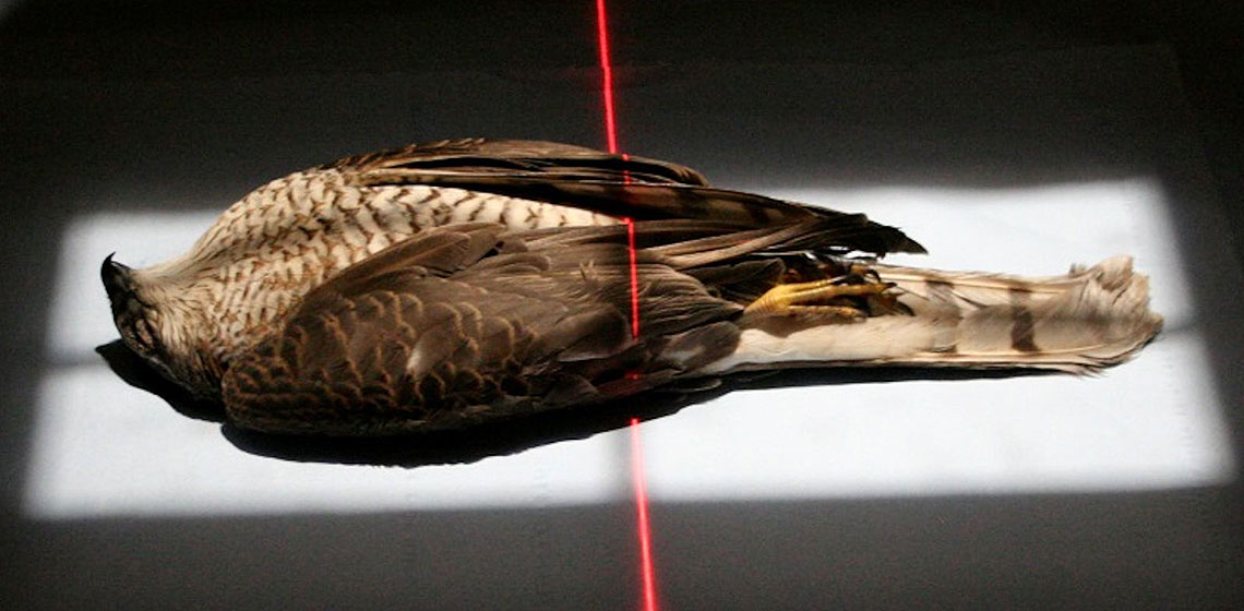
An immature, female Accipiter nisus (See Figure 6) was donated by the Natural History Museum Bird Group, Tring with the purpose of testing the above aims. The specimen named experimental mummy one (EM1) was presented to the Natural History Museum by a member of the public in February 2012 and was dry frozen upon receipt. It was transferred to a dry freezer and kept at -21 °C at the KNH Centre for Biomedical Egyptology, University of Manchester from October-November 2012. Over 72 hours, EM1 was thawed to room temperature and then radiographed (XR and CT) at the Manchester Royal Infirmary2 to ascertain any skeletal damage obtained peri-mortem (See Figure 7). Despite the cause of death being attributed to collision, no trauma was visible and no pathology was evident (See Figure 7). EM1 was then placed in a humidity controlled unit for 48 hours with an average temperature range of 45-48 °C in order to accelerate natural desiccation whilst simultaneously inhibiting bacterial growth (See Figure 8).
Mummification method
Firstly, EM1 was weighed to ascertain the body mass prior to mummification. The cadaver was placed upon a length of linen fabric3 to enable ease of manipulation and wrapping following the application of the pine resin:beeswax compound. The final resting position of the bird was maintained and was in the same position as those commonly seen in the ancient bird mummy bundles with the cranium in the normal anatomical position, the wings gathered around the upper body and the legs outstretched with a bend in the femoral-tibiotarsal joint (see Figure 9).
The beeswax and resin were weighed to ensure the same ratio as found in the Buckley et al. (2004) study (See Footnote 1). The ingredients were heated in a stainless steel pan until the compound had liquefied and the two materials had emulsified. This was then poured over the bird, with a particular focus on the chest and abdominal area (See Figure 10), after which the linen was folded to cover the total length of the bird to create the initial layer (See Figure 11).
The adhesive nature of the compound ensured that the linen was secured to the bird. There was a slight malodour during this process, most likely as a result of the heat of the emulsion being in direct contact with the cadaver. Several layers of linen were wrapped around the total body length of the bird, which resulted in a rectangular shape. The tension of the wrappings during this stage was relatively tight, although this increased with the concentric wrapping technique which followed. During the wrapping process, the resin:beeswax emulsion solidified and by the time the wrapping was complete, the bundle was solid to touch. This allowed the bird to be moved relatively rigorously to achieve the desired tension of the linen. The ends of the linen pieces were secured in place with small quantities of the resin:beeswax emulsion (See Figure 12).
Post-mummification observations
The mummy bundle was stored in a fume cupboard and the environmental conditions, both temperature and humidity, in addition to alterations in the appearance, odour and bundle mass, were recorded. Further radiography (XR and CT) was undertaken on three separate occasions post-mummification (at day 5, day 61 and day 229) to document the taphonomic changes.
Reduction in bundle mass: macroscopic and radiographic observations

Graph 1. Graph showing the gradual reduction in the mass of the bird bundle over the course of 220 days.
Immediately post-mummification (PM), the body mass of the mummy bundle almost doubled in size with the addition of the resin:beeswax emulsion and the linen. However, over the course of 220 days, the mummy bundle lost 133.54 g at a gradual rate (See Graph 1) and continues to reduce in mass. The sharp decrease and subsequent increase in bundle mass recorded over days 21-39 (December 2012-January 2013) (See Graph 1) was thought to be due to the change to in lab environmental conditions. Temperature regulation was stable, although humidity was recorded at 52 % on day 39. Throughout the experiment and with the exception of this period, the humidity was recorded as below the 50 % mark. Further understanding of the effects of humidity on the reduction in bundle mass requires investigation.
The potential reasons for this overall reduction in bundle mass (with the exception of the aforementioned point) were thought to have been attributed to two factors:
- The evaporation of the mummification materials; in this case the pine resin:beeswax emulsion. Interestingly, the radiodensity of ‘resin’ in the ancient Egyptian mummified animals was not directly reflected by this experiment. The resinous materials in the ancient specimens appear more radio-dense than in the experimental case. The degradation of the materials used by the ancient Egyptians in the mummification of animals requires further research.
- The reduction in size of the visceral and muscle tissues; this was clearly noticeable using CT images from pre- and post-mummification (see Figure 13). It appears that desiccation is continuous, even after the anointment and wrapping methods were completed.
Overall stability of the mummy bundle post-mummification
At the beginning of the post-mummification stage, the mummy bundle was malodorous in close contact, although this dissipated throughout the experiment. The solid nature of the mummy bundle noted during the mummification, a result of the solidifying resin: beeswax emulsion, was maintained. However, radiography showed that the linen wrapping method did not provide the amount of tension as was envisaged during the process, in particular around the tail region. The current state of the mummy bundle is stable.
The overarching result of this experiment proved that evisceration and desiccation through the use of natron was not required on small birds of prey to produce a well-preserved dead body.
Future Work
Further experimental mummification of bird subjects is required to continue to assess the replicability of ancient Egyptian materials and methods evidenced in archaeological research. The preservation of EM1 was achieved using a very simple method and represented only one species of bird known to have been mummified as a votive offering. Future work will continue using birds of prey which will gradually increase in size, in particular species identified from ancient mummy bundles, to understand how the size of bird cadavers may affect the mummification method by testing the hypothesis that large animals required evisceration to achieve preservation. In addition, further control of environmental conditions will be attempted (and recorded through the use of a sensor) by creating an enclosed dehydration/desiccation apparatus with a bed of Egyptian desert sand and an average temperature range of 25-30 °C.
Acknowledgements
The authors would like to thank Mrs P. Lusher for the donation of the bird to the Natural History Museum Bird Group, Tring and Dr. Joanne Cooper, Natural History Museum Bird Group, Tring, for donating the bird to this project. Thanks also go to Professor Andrew Chamberlain, KNH Centre for Biomedical Egyptology, University of Manchester for his advice and input into such research. In particular we are forever grateful to Professor Judith Adams and her radiography staff, Central Manchester University Teaching Hospitals NHS Foundation Trust, for the use of the imaging facilities and advice on the taphonomic changes.
- 1Resinous substances from a bird mummy, identified as a hawk by the museum and provenanced to Tarkhan in the 23rd-25th Dynasty (818-664 BC), were analysed using Gas Chromatography-Mass Spectrometry (GC-MS). The compound was composed of wax and fat/oil in 90:10 relative abundance (%), thought to have been beeswax and pine resin (Buckley et al. 2004, 295). The same relative abundance of these ingredients was employed in this research.
- 2XR parameters: Wrist AP and Lat 59.8kV, 2.1 mAs, focal spot size 0.6mm Siemens Medical System (Malvern, Pennsylvania, USA). CT parameters: 120 kV 11-13 mAs 0.625mm GE Lightspeed 32-row spiral MDCT (General Electric, Milwaukie, USA).
- 3Purchased at a fabric shop, UK. Measured 1 m in length and was cut into approximately 10 cm wide strips prior to mummification.
Country
- Egypt
- United Kingdom
Bibliography
ARMITAGE, P. L. and CLUTTON-BROCK, J. 1981. “A radiological and histological investigation into the mummification of cats from ancient Egypt” Journal of Archaeological Science: 185-196
ATHERTON, S. D. 2012. An investigation of the post-mortem status and mummification practices of avian votive mummies in ancient Egypt. University of Manchester, unpublished thesis.
CLIFFORD, W. and WETHERBEE, M. 2004. “Making a duck mummy” KMT A modern journal of Ancient Egypt, pp. 64-67.
COCKBURN, A., COCKBURN, E. and REYMAN, T. A. 1998. Mummies, Disease and Ancient Cultures (Second Edition). Cambridge. Cambridge University Press.
GARNER, R. 1979. “Experimental Mummification” in David, A. R. (ed.) The Manchester Museum Mummy Project: multidisciplinary research on ancient Egypt mummified remains. Manchester. Manchester University Press.
IKRAM, S. and DODSON, A. 1998. The Mummy in Ancient Egypt: equipping the dead for eternity. London. Thames and Hudson Ltd.
TAYLOR, J. 2001. Death and the Afterlife in Ancient Egypt. London. The British Museum Press.
IKRAM, S. and ISKANDER, N. 2002. Catalogue Général of Egyptian Antiquities in the Cairo Museum: Nos. 24048-24056; 29504-29903 (selected); 51084-51101; 61089 Non-human mummies. Cairo. The Supreme Council of Antiquities Press.
IKRAM, S. 2005. Divine Creatures: animal mummies in ancient Egypt. Cairo. American University in Cairo Press.
LUCAS, A. 1962. Ancient Egyptian Materials and Industries. Fourth Edition revised by J. R. Harris. London: Edward Arnold Publishers.
MCKNIGHT, L. M. 2010. Imaging applied to animal mummification in ancient Egypt. BAR International Series 2175.
MCKNIGHT, L. M. and ATHERTON, S. D. 2014 “How to pigeon-hole your mummy – a proposed categorisation system for ancient Egyptian wrapped animal remains based on radiographic evaluation” Yearbook of Mummy Studies 2, 109-116
KESSLER, D. and NUR EL-DIN, A. H. 2005. “Tuna al-Gebel: millions of ibises and other animals” In Ikram, S. (ed.) Divine Creatures: animal mummies in ancient Egypt. Cairo. American University in Cairo Press.
RAY, J. D. 2011. Texts from the baboon and falcon galleries: Demotic, Hieroglyphic and Greek inscriptions from the Sacred Animal Necropolis, north Saqqara. London: Egypt Exploration Society.
Von den DRIESCH, A., et al. 2006. “Mummified, deified and buried at Hermopolis Magna – the Sacred Ibis from Tuna el-Gebel, Middle Egypt” Ägypten und Levante 15: 203-244.
VOS, R. L. 1992. The Apis Embalming Ritual. Leuven: Peeters.

